Histiotic Sarcoma
Definition
Localized histiocytic sarcoma, disseminated histiocytic sarcoma and malignant histiocytosis are fairly rare tumors overall but occur with high incidence in Bernese Mountain dogs, Rottweilers, Flat Coated Retrievers, and Golden Retrievers. Histiocytic sarcomas are very aggressive tumors, and can therefore become very invasive (destroy normal surrounding tissues) as well as have a high rate of metastasis (spreading to other areas of the body). Localized histiocytic sarcoma lesions most commonly are found in the spleen, lymph nodes, lung, bone marrow, skin, brain, and joints of the limbs. Disseminated histiocytic sarcoma and malignant histiocytosis are multi-system, rapidly progressive diseases in which there is simultaneous involvement of multiple organs such as the spleen, lymph nodes, lung, bone marrow, and skin.
What are the common signs of this cancer?
Dogs with histiocytic sarcoma typically have non-specific signs, such as anorexia, weight loss, and decreased energy. Other signs depend on the organs involved and are usually a consequence of destructive mass formation. For example, if there is a large mass in the lungs, a dog may experience coughing or difficulty breathing. If the brain is involved, you may see seizures, incoordination, and paralysis. If a joint is involved, lameness or limping is often seen. Read more
Lung Cancer
Definition
The lung is the essential respiration organ whose principal function is to transport oxygen from the atmosphere into the bloodstream, and to release carbon dioxide from the bloodstream into the atmosphere. There are two type of lung cancer diagnosed in dogs. The first is primary lung cancer which is defined as lung tumors that originate in the lung tissue. The second type is metastatic lung cancer whichis cancer that originates elsewhere in the body such as a leg bone, the mouth, or the thyroid gland, but has spread to the lung via the bloodstream.
Primary lung cancer, or tumors originating in the lung, are relatively uncommon in dogs (less than 1% of all cancers in dogs), although the number has been increasing. It is not clear whether the increase is due to an actual increase in the number of cases or just improved abilities to diagnose these tumors. Primary lung tumors are almost always malignant and are usually carcinomas (often adenocarnimas), although can be hemangiosarcomas. They usually present as a large solitary mass visible in the lung on a chest x-ray.
Carcinomas develop from the epithelial tissues in the lungs. They may be primarily derived from the lung tissue itself, or the airways or bronchioles. Canine lung cancer is aggressive and frequently metastasizes to the regional lymph nodes and tissues of the thorax. Adenocarcinoma has a tendency to metastasize (spread) to the central nervous system. Adenocarcinomas are further classified based on their location (eg bronchial, bronchoalveolar, or alveolar carcinoma).
A classification system based on the prevailing histologic pattern may be used to categorize most primary lung neoplasms as adenocarcinomas, squamous cell carcinomas, anaplastic carcinomas, or bronchioloalveolar carcinomas. Only the neoplastic cell type and the presence or absence of metastatic disease have been found to be of value as prognostic indicators.
Metastatic lung tumors are those that spread from a primary tumor elsewhere in the body. The lungs are a common site to which other types of cancer spread, such as with dogs diagnosed with bone cancer. Metastatic lung tumors are usually found in multiples, not as a single mass. Be aware that dogs with multiple lung masses may be the result of a fungal infection and not cancerous. That is why it’s imperative to obtain an accurate diagnosis if lung masses are found in your dog. Read more
Brain Cancer
Definition
A brain tumor is any intracranial tumor created by abnormal and uncontrolled cell division, normally either found in the brain itself, in the cranial nerves, in the brain envelopes (meninges), skull, or pituitary and pineal gland. Primary brain tumors (those arising form the cells of the brain and it’s lining) in dogs include meningioma, glioma, choroid plexus papilloma, pituitary adenoma or adenocarcinoma, and others.
Secondary or metastatic brain tumors originate from malignant tumors (cancers) located primarily in other organs and metastasize (spread) to the brain. These include hemangiosarcoma, mammary carcinoma and melanoma. These tumors carry a very poor prognosis because they have already spread through the body.
Brain tumors are not uncommon in older dogs, however there is an increasing incidence of brain tumors occurring in younger dogs. Brain tumors vary widely in their level of malignancy and some can be treated quite effectively. The most common forms of brain tumors are outlined below. Read more
Abdominal Cancer
Cancer in the Stomach
Stomach cancer can be a silent killer because a dog is usually in the advanced stages by the time he shows signs of illness. Learn about the different types of cancer that typically affect the stomach, as well as the symptoms a sick dog might display.
Canine stomach cancer is rather rare. It accounts for about 0.1 percent (1 in 1000) of all cancers in dogs. However, certain breeds seem to be more predisposed to stomach cancer. For example, Chow Chows have between 10 to 20 times the risk of stomach cancer compared to other breeds. Read more
Canine stomach cancer is rather rare. It accounts for about 0.1 percent (1 in 1000) of all cancers in dogs. However, certain breeds seem to be more predisposed to stomach cancer. For example, Chow Chows have between 10 to 20 times the risk of stomach cancer compared to other breeds. Read more
Gastrointestinal Cancer
You might have no clue until it’s too late, but constant worry is the real terminal prognosis. Canine gastrointestinal cancer is a gruesome, silent killer, with dogs only showing symptoms when the disease has reached advanced stages.
Don’t freak out; canine gastrointestinal cancer is not at all common. It accounts for only 1 out of every 1000 cases of canine cancer. Nevertheless, you should stay vigilant and keep your dog healthy by feeding them a healthy diet, exercising often, and getting regular health checkups, especially if you have a breed like a Chow Chow, Akita, or Keeshond, among others.
The most common cause of stomach cancer in dogs is adenocarcinoma, a tumor of the glandular tissue that grows to reach the stomach and other vital organs. Read more for an extensive breakdown of different types of gastrointestinal cancer in dogs and their tell-tale signs.
Lymphoma, LSA
WHAT IS CANINE LYMPHOSARCOMA (LYMPHOMA, LSA)?
Lymphoma is one of the most common cancers of dogs. It is a cancer that derives from the cells in the lymph system, which is comprised of the lymph nodes and circulating lymphocytes in the vascular system. Although lymphoma can affect virtually any organ in the body, it most commonly arises in organs that function as part of the immune system such as the lymph nodes, spleen, and bone marrow. By far the most common type of lymphoma in the dog is multicentric lymphoma, in which the cancer is usually first noticed in lymph nodes. Lymphoma is also one of the most well understood cancers in dogs, with regard to diagnosis, treatment options, and prognosis. It is one of the few cancers in dogs that can have remission times of years, and although it is not frequent, a cure is sometimes a possibility.
THE CAUSES OF CANINE LYMPHOMA
The cause of lymphoma is not well understood. Certain breeds of dogs, such as Golden Retrievers, are at higher risk of getting lymphoma. Therefore, it is believed that there is a significant genetic component to lymphoma. However, any breed of dog, at any age, including juvenile dogs, can get lymphoma. It is also believed that there are environmental contributors to lymphoma. Environmental factors such as chemicals, airborne pollutants, and other variables may damage cells. As dogs age, cellular repair is slower, and cancer becomes more prevalent. Other than protecting our dogs from known predispositions or causes of cancer such as secondhand smoke, or certain chemicals, we really cannot prevent our dogs from getting lymphoma. Read More.
Mast Cell Tumors
WHAT ARE CANINE MAST CELL TUMORS?
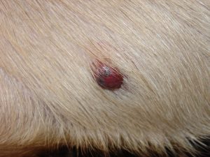
Canine mast cell tumors (MCTs) are a common malignant tumor in dogs. Mast cell tumors are regulators of the immune system cells. While MCTs can be found in many organs of the body, it is the most common malignant skin tumor in dogs.
The normal mast cells are important in inflammation and allergic reactions. The MCTs themselves however, are formed by many trillions of malignant mast cells. They become malignant by mutating in a way that allows them to escape the natural process of cell aging and death. For that reason they are able to grow unchecked, and these tumors can range from relatively benign to being very aggressive.
Malignant mass cells are also not as stable as the normal mast cells. Each cell contains many substances that can cause bruising, bleeding, swelling, and inflammation. Any trauma directly to the tumor such as surgery, scratching, bumping, or crushing the tumor may cause release of inflammatory mediators. In this situation the patient may display symptoms of an allergic reaction without any contact with allergens or parasites.
THE CAUSES OF CANINE MAST CELL TUMORS
It is hard to find a definitive cause, but the thought is that genetics plays a part in this type of cancer. There are certain breeds that can be more affected, such as Labrador Retrievers, Golden Retrievers, Bulldogs, Boxers, Boston Terriers, Cocker Spaniels, Schnauzers, and Shar-peis. Read more
Hemangiosarcoma
WHAT IS CANINE HEMANGIOSARCOMA?

|
Canine hemangiosarcoma or HSA is cancer that develops in the cells that form blood vessels (endothelial cells). Interestingly, dogs are the species that are the most diagnosed with this type of cancer. It is estimated that as many as two million dogs get this cancer. Most will, unfortunately, die from HSA, as the disease is usually incurable. There is one form of HSA that is generally less aggressive, and that is cutaneous melanoma. The cause has been attributed to exposure to the sun and is generally treatable with surgery.
Dogs are usually middle-aged and older when they develop the disease, and some breeds are known to be predisposed to HSA. German Shepherds, Golden Retrievers, Portuguese Water Dogs, and Skye Terriers are four of them. That being said, any breed and sex can develop HSA. |
THE CAUSES OF CANINE HEMANGIOSARCOMA
|
Much more research is needed for this type of canine cancer. Sun exposure and genetics appear to result in an increased risk of developing this tumor. The exact cause is unknown.
|
THE CAUSES OF CANINE HEMANGIOSARCOMA
|
SYMPTOMS OF CANINE HEMANGIOSARCOMA

|
The biggest problem with this disease is that the symptoms can mimic a benign tumor such as a hematoma, or an inability to clot blood, such as seen when a dog ingests rat poison. Quite often you do not even know the tumors are present until a bigger problem presents itself, such as blood loss from a ruptured spleen or liver. They are frequently painless as well, which makes detection of an issue difficult.
Clinical signs that the family might notice include pale gums, rapid breathing, and acute weight gain due to fluid, often blood, in the abdomen, extreme fatigue or lethargy, and eventual collapse. These tumors can grow and then rupture. Since the tumors develop in the cells that form blood vessels, the ruptures cause acute bleeding. That brings on the symptoms mentioned above here. While blood is not seen outside of the dogs’ body, a sick dog is losing blood internally. |
DETECTION AND STAGING
The staging generally consists of a minimum of blood work, three-view chest X-rays, abdominal and heart ultrasound, and urinalysis. There may be other tests ordered, or the animal may arrive in a state of shock due to internal bleeding, necessitating immediate supportive care prior to surgery; IV fluids, pressure wraps to help stop bleeding, and blood pressure checks, for example.
Surgery is usually recommended, mainly because there is really no other way to see for sure if a mass is malignant or not. It is also the only way to stop a patient’s internal bleeding. This not only can save the dogs’ life at this particular time but a tissue sample can be taken to confirm a diagnosis of HSA.
Surgery is usually recommended, mainly because there is really no other way to see for sure if a mass is malignant or not. It is also the only way to stop a patient’s internal bleeding. This not only can save the dogs’ life at this particular time but a tissue sample can be taken to confirm a diagnosis of HSA.
TREATMENT
|
Hemagiosarcomas can occur anywhere in the body and may present as a mass on sites such as the spleen, liver, muscle, heart, under the skin, on the tongue, and on the skin. Surgery is the single most effective treatment for patients with hemangiosarcomas. Unfortunately, for most patients, it is not possible to determine the cause of the bleeding until the mass is evaluated by a pathologist after it was removed. For example, for dogs with a mass on the spleen, 2/3rds of them are malignant, one third is not. Of the two-thirds of the dogs with a malignant splenic mass, two-thirds of them have hemangiosarcoma. To make things more difficult, not all hemangiosarcomas are the same; some are low grade with a more favorable prognosis, whereas others are intermediate and high-grade hemangiosarcomas. The high-grade tumors are generally quite aggressive.
Thus, even though surgery may be essential to save the dogs’ life in a crisis, it is rarely the only answer. Since hemangiosarcoma is a very aggressive disease, the life expectancy after surgery maybe a few weeks to a few months. Chemotherapy is generally recommended after surgery since this cancer metastasizes early and quickly. doxorubicin-based chemotherapy is used most commonly. |

|
FINALLY
The average survival time for dogs with a malignant hemangiosarcoma after both surgery and chemotherapy may be only six to eight months. A great deal of research is underway to find new treatments for this very aggressive disease.
Osteosarcoma
WHAT IS CANINE OSTEOSARCOMA?
Osteosarcoma is the most common type of bone cancer in dogs. It can occur anywhere on the skeleton, however it is most commonly diagnosed at the end of the bones (metaphysis) of the legs. This cancer has many similarities to osteosarcoma in children. It is painful in both kids and dogs. It almost always metastasizes (spread) to other organs, especially the lungs and other bones. It is said that up to 85% of tumors originating in the skeletal system is osteosarcoma. Osteosarcoma as a rule becomes evident in the later years when a dog is between the ages of 7 to ten.
THE CAUSES OF CANINE OSTEOSARCOMA
Canine osteosarcoma occurs most commonly in large or giant breeds, such as Great Danes, Scottish Deerhounds, Rottweilers, and Greyhounds. The risk also seems to increase when young dogs are fed diets that promote rapid growth. Read more
Mammary Carcinoma
WHAT IS CANINE MAMMARY CARCINOMA?
Breast cancer in female dogs and in women is common. The disease in dogs is more commonly known as canine mammary carcinoma or mammary gland adenocarcinoma. This tumor is the most common cause of death and the most common cancer in the unsprayed dog. The tumor is also quite common in dogs that were spayed late in life because exposure to female hormones early in life increases the prevalence of this tumor. About half of mammary tumors are malignant and about half of the malignant tumors have metastasized at the time they are first diagnosed. The great news is that most dogs with breast tumors are cured with appropriate surgery.
THE CAUSES OF CANINE MAMMARY CARCINOMA
As mentioned above, it is the non-spayed female that is the most likely to develop this cancer. This is especially true as the dog ages. Of the dog is spayed before her first heat (estrus) the risk of canine mammary carcinoma is only 0.05% – very low. After the first heat cycle it goes to 8%, and climbs dramatically to 26% if spayed after the second estrus. In contrast, male dogs are only at a 1% or less risk for this cancer.
If a dog is spayed after initial diagnosis, some studies show that the survival rate is higher than those who remain intact after diagnosis. Read more
If a dog is spayed after initial diagnosis, some studies show that the survival rate is higher than those who remain intact after diagnosis. Read more
